In this complete guide to CL in goats, we answer the most common questions that farmers ask about caseous lymphadenitis in goats.
Caseous Lymphadenitis (CL) is a very serious, contagious condition in goats. ALWAYS consult with your veterinarian and take appropriate precautions when dealing with a goat suffering with CL.
Table of contents
What is CL in Goats?
Caseous Lymphadenitis is a bacterial infection of lymph nodes of goats. This disease is characterized by wounds in the outer lymph nodes of the abdomen and neck region. However, these abscesses can develop inside the lung, liver, udder, and kidney.
The Goats having CL can cause financial losses because of goat’s death, decrease in the production of the milk, and reduction in weight of the animal. Additionally, farmers that have flock free of CL will avoid buying animals from the farm that has disease outbreak as it will reduce the worth of breeding herd. Slaughtered goat hides may also be worthless due to the outbreak of this viral disease.
Importance of Lymph Nodes
Lymph nodes have a crucial function in the immunity of the animals. They store blood cells and filter foreign things to detect the infection. There are several lymph nodes in entire the body.
CL in Goats Symptoms
The goat gets the infection with the entrance of bacteria through the wound or membranes of nose, eyes, or mouth. The swelling of the lymph nodes are not visible for a few months after the initial contamination. Mostly, the initial abscess develops in the neck and head region.
The most susceptible area for the injury is head such as combat wounds, a splinter from the wooden feeders etc. The infection will increase if the goat places the wounded head on the area where the other infected goat places the head.
As the wound in the lymph nodes grows, the abscesses containing pasty pus enriching with bacteria will develop. These abscesses dislocate the healthy tissue and can lead to problem in breathing, ruminating, eating, depending on the site of the abscess.
If the location of the abscesses is in the lungs, goats often display signs and symptoms of respiratory pain, which will decrease production and can cause death of the animal.
Transmission of CL in Goats
As the size of abscesses increases, they will rupture and the infection will direct transmit to the other goats of the herd. The discharge from the nose can be another source of transmission of the disease as lung lesion grows, rupture and then expel the contents.
Indirect transmission is another way of disease transmission through the infected types of utensils and ground in the shed. The goat infected with this disease must be milked at the end, and all the utensils must undergo proper cleaning and sanitization after the use.
CL Locations on Goats

External Abscesses
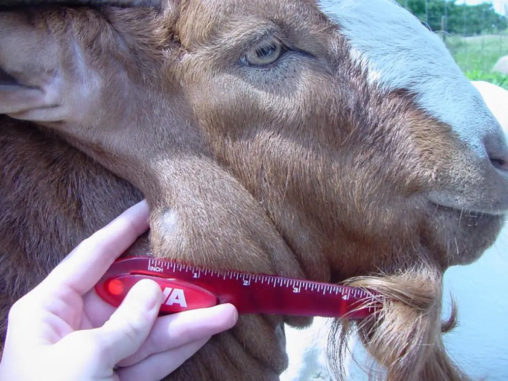
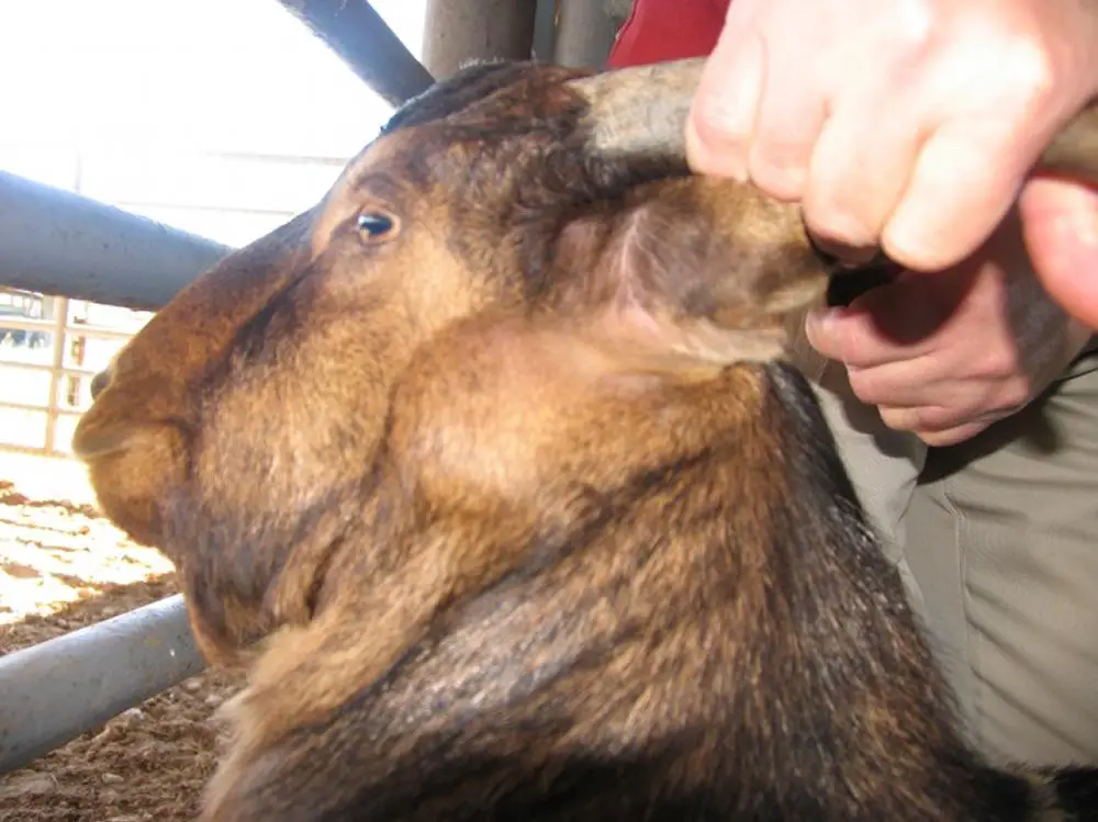
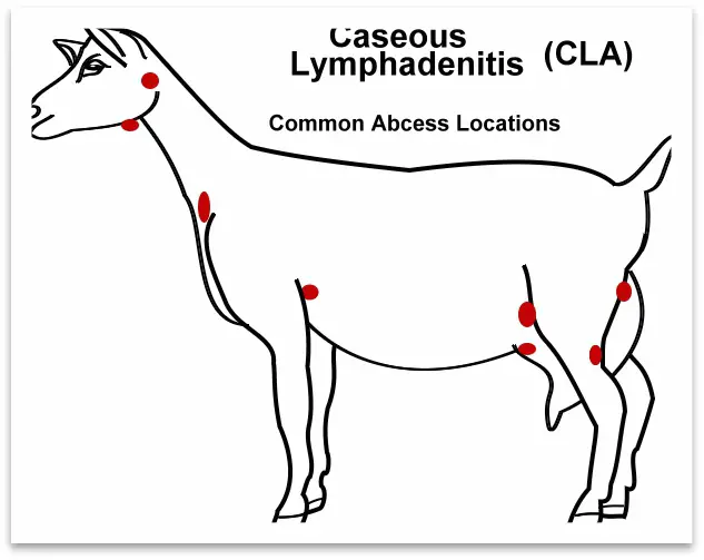
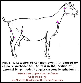
The CL often present as abscesses that are at the back of the ears, under the neck or jaw, on the shoulders, and seldom behind the legs where the udder or scrotum attaches. However, abscesses can arise anywhere on the body. These abscesses are frequently but not always adjacent to the lymph nodes.
The abscesses are soft when palpated. Some ulcers are well defined and rounded and contain thick white and yellow-greenish pus. The pus of these abscesses are odourless but can have strong smells in advanced sores.
Internal Abscesses
Internally, the abscesses develop on the organs and lymph nodes of the goats. Notably, organs impacted may include lungs, liver, and kidney. Theses abscesses can be seen during the necropsy.
Internal abscesses are fatal to the goats and generally cause weight loss. In CL, sores are more often present externally as compared to internally. It may take 2-6 months for physical signs to show after infection.
CL Testing in Goats
The diagnosis is normally done by observing a solid abscess at the site of the lymph node. The records of CL in a herd are sufficient to presume the diagnosis. The suspected goat should keep separate and must get the treatment as transmittable until the reason of the inflammation is identified.
Clinical signs and symptoms found by observation and by physical assessment will help to diagnose the disease. CL abscesses contain pus with a bad smell. X-ray, a biopsy, or postmortem examinations are the only method to observed the internal wounds.
CL in Goats Treatment
The infected animals will remain ill permanently, and CL will not respond to most of the antibiotics. Several studies conducted to see the appropriate medicine, its dosage, or extraction time of several antibiotics used to control this disease in goats is not obtainable. Therefore there is no particular treatment for CL.
The usage of antibiotics with no response will enhance the emerging issue of antibiotics resistance along with the wastage of time and wealth.
It is possible to drain the abscess surgically. However, this draining may cause the contamination of the surroundings, demanding the animals to isolate until the complete healing of the abscess. The background requires be properly cleaning and disinfecting to avoid the spread of the disease.
Surgical elimination of abscesses is not the practical solution for most farmers, but it eradicate the abscess without cleaning it so the bacterial don’t get into the surroundings. However, this requires to anaesthetized (unconsciousness) the goat for such a delicate procedure.
Control and Eradication of CL in Goats
The vaccine against CL for goat is accessible in the US. However, sheep vaccine for CL is available in Canada, although it is not advisable to use the sheep vaccine in goats due to the higher incidence of undesirable problems like fever, abortion, and inflammation at the injection site.
The best control from CL is to have a closed herd or allow the new animals to a strict separation protocol to avoid the infection from incoming to the herd. If the CL is already there in the herd, then the control of the disease can be hard.
A firm testing of all the goats can be helpful. It is advisable to manage the young ones by preventing their contact with the CL positive female goats and providing the young one’s heat-treated colostrums or pasteurized milk to avoid the spread.
Since the bacterial of CL survive for months in the environments, anticipation and disinfection policies are the key to control the transmission of the infection. Although the abscesses are noticeable, many goats that only have wounds internally go unobserved. Therefore, monitoring the signs and blood samples of goats will help to detect the infected goats.
CL in Goats – Contagious to Humans?
Infected goats are significant sources of contamination of CL in humans. Humans become infected by having direct contact with the diseased goat or by having the skin contact with pus from the infected goat. The signs of CL in humans include the development of painful skin wounds with pus and dead cells.
The disease is potentially transmissible to humans, so humans must wear proper protective clothing when working with the infected or possibly infected animals. This protection will help both humans and animals to remain safe from the infection.
CL in Goats – Pictures
Video of Veterinarian Treating CL Abscess
Summary
Even though CL is not a major reason of death in goats, production failure and fear for animal wellbeing should give confidence to producers to decrease the incidence of the outbreak. It is the accountability of the producers to prohibit CL entering into the herd by following proper bio-security and testing all the new animals. Farmers can control and eliminating CL from the herd with appropriate management and separating both infected and healthy herds.


